PT leads to improvements for patients with common jaw disorder

More research is needed that evaluates how effective these treatments are

Temporomandibular disorder (TMD) is a term used to describe a set of common conditions that affect the temporomandibular joint (TMJ). The TMJ connects the jaw to the skull and allows it to move up and down and from side to side. Typical symptoms of TMDs include pain (especially while chewing), difficulty opening the mouth, the jaw getting stuck, and a clicking or popping sound, all of which can have a negative impact on patients’ lives. There are a number of available treatments for TMDs, two of which are physical therapy and occlusal splints. Physical therapy usually consists of a variety of interventions that are all intended to reduce pain and improve strength, mobility, coordination, posture, and flexibility. An occlusal splint is a specific type of mouth guard that prevents clenching the jaw and protects the teeth from other harmful habits. While both of these treatments are commonly used for treating TMDs, there is no research that compares the two of them. For this reason, a powerful study called a randomized-controlled trial (RCT) was conducted to determine if physical therapy or wearing a splint is more effective for patients with a TMD.

Both interventions last for six weeks

To conduct the study, researchers recruited patients who were diagnosed with a TMD and screened them using specific criteria to determine if they could participate. From this search, 112 individuals were accepted to the study and randomly assigned to either the physical therapy group or the splint group. The physical therapy treatment program took place during three 15-minute sessions per week for six weeks. During these sessions, a physical therapist led patients through a series of exercises and stretches to relax the jaw. In the relaxed jaw position, for example, patients were told to place their tongue behind their upper front teeth and allow the teeth to come apart in order to relax the jaw muscles. In the splinting group, patients were given an occlusive splint and told to wear it every day for the next six weeks as prescribed by a dentist. Measurements of pain levels and jaw flexibility were taken for all patients at the start of the study and then again at the end of the interventions six weeks later.

Patients with a TMD should see a physical therapist to receive similar treatments

When patients were assessed at the end of the study, it was found that those who followed a course of physical therapy experienced significantly greater improvements compared to those who wore a splint. This was found to be the case for both pain levels and flexibility of the jaw, the two outcomes that were measured. These results suggest that physical therapy provides superior outcomes for patients with a TMD than wearing an occlusal splint. Physical therapy has also been found to be a safe, relatively easy, and inexpensive intervention, which makes it even more attractive. For these reasons, if you currently have a TMD and are bothered by pain, it’s recommended that you see a physical therapist. Doing so will get you on the right track and provide you with the treatment needed to manage your symptoms and regain the ability to move your jaw without pain.

-As reported in the September ’18 issue of The Journal of Physical Therapy Science

You can alleviate your jaw pain with some simple home remedies

Whether you realize it or not, you use your temporomandibular joint (TMJ) pretty often. This joint connects your lower jaw to the rest of the skull right in front of your ears, and you can actually feel it if you touch right below your temples and open your mouth. The TMJ allows the jaw to move up and down and from side to side, which is why it’s used with practically every movement that involves your mouth. So when you speak, swallow, or bite down on something, you have both of your TMJs to thank.

Unfortunately, the TMJ also has a reputation of being a common location for pain. Temporomandibular disorder (TMD) is a general term used to describe any condition that affects the TMJ. The common trait of all TMDs is pain and possibly inflammation in the muscles of the jaw and surrounding area. This pain can spread to the cheek, ear, or temple, and it often causes difficulty performing any tasks that require opening or closing the mouth. As with many other parts of the body, you might not become acquainted with your TMJ until something goes wrong with it, as is the case with these conditions.

Approximately 5-12% of the population is affected by a TMD, with the majority of these individuals dealing with acute pain—meaning it developed recently. If you count yourself as one of the millions of Americans dealing with a TMD right now, you may be curious if there are any simple solutions out there that will help to alleviate your symptoms. The good news is yes, there are. Most cases of acute pain from a TMD can be effectively treated with some home remedies that you can perform on your own. We recommend the following tips for a recent onset of pain or other symptoms to the TMJ:

  • Heat therapy: applying heat with a moist warm towel or dry heating pad can reduce jaw pain and stiffness by increasing the flow of blood to the area; it’s recommended that you use a warm compress and place it on the jaw and temples for about 10-15 minutes, about twice a day; heat therapy is particularly effective if you have a dull, steady, aching pain
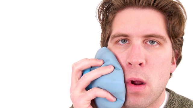
  • Ice therapy: if the heat does not lead to any notable improvements after two days, try applying ice instead; the application of ice will numb the nerves and dull the pain by slowing down the flow of blood to the area; apply an icepack wrapped in a towel to the area for about 15-20 minutes a few times a day
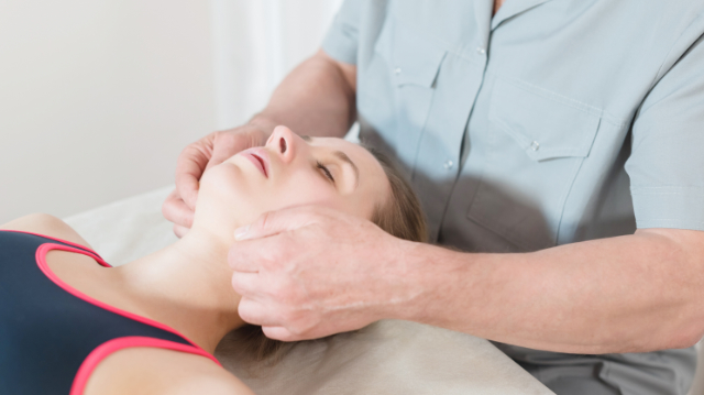
  • Massage: massaging the areas around your jaws can also improve blood flow and provide relief; this can be accomplished by opening your mouth and locating the muscles next to your ears by the TMJ; put your fingers on any area that’s sore and apply gentle pressure in a circular motion; you can also massage the muscles on the sides of your neck if there is tension in that region as well
  • Jaw exercises: regularly moving the jaw in a specific and systematic manner may be helpful for further reducing TMD symptoms; a selection of exercises is below
    • Relaxed jaw exercise: rest your tongue gently on the top of your mouth behind your upper front teeth; allow your teeth to come apart while relaxing your jaw muscles
    • Chin tucks: with your shoulders back and chest up, pull your chin straight back, creating a “double chin;” hold for three seconds and repeat 10 times
    • Resisted opening of the mouth: place your thumb under your chin; open your mouth slowly, pushing gently against your chin for resistance; hold for three to six seconds, and then close your mouth slowly
    • Resisted closing of the mouth: squeeze your chin with your index and thumb with one hand; close your mouth as you place gently pressure on your chin; this will help strengthen your muscles that help you chew
  • Pain-relieving medications: non-steroidal anti-inflammatory drugs (NSAIDs) like ibuprofen and other over-the-counter pain medications may help to reduce inflammation and alleviate pain in some individuals
  • Splint or night guard: these devices fit over your upper and lower teeth to prevent these rows from touching one another; in effect, this will lessen the impact of clenching or grinding the teeth and correct your bite by putting your teeth in a better position

Avoid a jaw problem by chewing properly and practicing good posture

There are over 200 bones in the human body, and the head and face account for 22 of these. Of the face and head bones, the lower jawbone—or mandible—is the only one that can move. The mandible is connected to the temporal bone of the skull at two points just in front of each ear through an important joint called the temporomandibular joint. This joint gets lots of attention not only because it allows us to move our jaw in order to talk and chew food, but also because it’s a common location for pain.

A temporomandibular disorder (TMD) is essentially any condition in which the temporomandibular joint is not functioning properly. These types of issues are quite common, as they affect millions of Americans every year and can strike at any age. Symptoms vary from patient to patient, but in most cases they lead to pain and discomfort in the jaw and surrounding facial muscles which can make it difficult to perform any actions that require opening or closing the mouth.

It’s not entirely clear what causes TMDs, but several factors may be responsible, including injury to the jaw, repeated clenching or grinding of the teeth, high stress levels, muscle spasms, and arthritis. This uncertainty, combined with the fact that we need to use our jaws constantly, might lead you to believe that there’s not much that can be done to avoid a TMD. Fortunately this is not the case, as there are several steps you can take to reduce your risk for TMDs. We recommend the following strategies:

  • Be more careful with what and how you chew
    • Avoid chewing gum
    • Chew with both sides of your mouth
    • Avoid eating too many hard or crunchy foods
    • Take smaller bites of food
    • Don’t bite on hard objects like pens or pencils
    • Avoid biting your nails
  • Practice good posture
    • Keep your head balanced and not hunched forward, your shoulders straight, and torso in alignment with your head and shoulders (with good posture, a straight line can be drawn from your ears to your shoulders)
    • Avoid cradling your phone between your neck and shoulders
    • Try not to regularly carry a heavy purse or backpack on one shoulder
    • Consider using ergonomically-designed products for your office setup
  • If you grind or clench your teeth, try to reduce or stop this habit
  • Try to keep your tongue at the roof of your mouth and avoid letting your teeth touch; your teeth should be kept at least a few millimeters apart unless you’re chewing, and even then they should not be making much contact with one another
  • Sleep on your back or side and avoid sleeping on your stomach, which can strain your jaw; make sure your pillow provides enough support for your head and neck
  • If your stress levels are high, explore options to reduce your stress, including meditation, yoga, mindfulness practices, and cognitive behavioral therapy
  • Don’t rest your chin in your hands
  • Breathe through your nose and keep your lips together

Jaw disorders can cause problems for similar reasons at different ages

Pain can develop just about anywhere in your body, and regardless of where it occurs, can prove to be an annoying problem. The jaw is one region that doesn’t get discussed as often as more common injury sites like the knees or the neck, but disorders of the jaw are actually more prevalent than you may think. These types of issues—which are called temporomandibular disorders—affect millions of Americans every year and typically cause nagging symptoms like jaw pain and headaches. Some people are more likely than others to have a problem with their jaw, but they can occur in anyone and at any age.

The temporomandibular joint (TMJ) is a hinge joint that connects the part of the skull directly in front of the ears (temporal bone) to the lower jaw (mandible). It allows you to move your jaw up and down and from one side to the other, which is necessary for talking and chewing. Temporomandibular disorder, or TMD, is a general term used to describe a variety of conditions that cause pain and dysfunction in the jaw, jaw joint, and surrounding facial muscles that control chewing and jaw movement.

The definite cause of TMDs is still unclear, but some theories suggest that they may be due to injury in that region, grinding, or clenching teeth, osteoarthritis, or stress. Symptoms vary from patient to patient, but some of the more common signs of a TMD include the following:

  • Jaw pain or tenderness, which can be on one or both sides of the jaw
  • Aching pain in/around your ear or in your face
  • Difficulty opening/closing the mouth or chewing
  • Popping, clicking, or locking of the jaw
  • Headaches
  • Ear aches or ringing in the ear

TMDs can occur as early as infancy, but are not frequently seen in this age group and are more likely to develop somewhat later in life. When a newborn or young child is diagnosed with a TMD, it’s typically related to a deformity that was present at birth. But as children age, the likelihood of experiencing a TMD increases, particularly around the teenage years. Girls are more likely than boys to have an issue with their jaw.

It’s difficult to define exactly what causes a TMD in children and adolescents, but most experts believe that overexerting the jaw is at least partially responsible. Overexertion can occur from high levels of stress or anxiety, repeatedly clenching the jaw or grinding teeth—which is called bruxism—or from a traumatic injury such as one sustained in sports. High stress levels can actually make kids more likely to tighten their jaws, and over time, these behaviors will change the alignment of one’s bite and affect the muscles used for chewing. Regularly using a forward head posture with slouched shoulders and a rounded upper back may also contribute to the development of TMDs, since this type of posture can affect how the jaw closes.

The risk for TMDs continues to increase to a certain point, as the majority of cases are seen between the ages of 20-40. About 15% of adults currently suffer from a TMD, and once again, women are at least twice as likely to get them than men. As with younger populations, a highly stressful lifestyle, injuries to the jaw, and frequent jaw clenching will all remain factors that could play a role in the development of a jaw problem. Unregulated stress and a long-term habit of jaw clenching will likely increase this risk even more on account of the repeated stress placed on the jaw over time.

But there are also some risk factors that are unique to adults and may also increase the risk for developing a TMD. Arthritis—which includes rheumatoid arthritis, osteoarthritis, and gouty arthritis—is more likely to occur later in life. These types of arthritis can affect various joints of the body, and if one of the TMJs is involved, the risk for experiencing a TMD may be higher. Muscle spasms caused by other medical conditions or injuries may, as well as changes in the structure of teeth from dental procedures or the wearing down of teeth may also become more likely in older age. Both of these factors can also play a part in the development of a TMD.

Value of PT is found to be far better than surgery for meniscus tears

Understanding the costs associated with each treatment will help doctors and patients make better decisions

The meniscus is a wedge-shaped piece of cartilage between the thighbone and the shinbone. There are two menisci (plural of meniscus) in each of your knees, and the job of each of these structures is to stabilize the knee joint and absorb shock. Tears of the meniscus are very common, and many of these injuries are treated with a surgical procedure called arthroscopic partial meniscectomy (APM). This procedure involves a surgeon using small incisions to guide a camera and surgical instruments to remove part of the meniscus, and it’s currently one of the most commonly performed surgeries in the world. Millions of arthroscopic knee surgeries are performed throughout the world each year, and even though it’s not clear if surgery leads to better outcomes than conservative (non-surgical) treatment, the number of these procedures is decreasing slower than expected. One way to change this trend is by evaluating the costs of surgery compared to conservative treatments like physical therapy to help doctors and patients make better decisions, but no study of this nature has been performed yet. Therefore, a study was conducted that evaluated the costs of physical therapy versus APM for patients with meniscus tears to determine which of the two treatments is more cost-effective for patients.

Large group of patients assessed for two years

Data was collected from an ongoing study called a randomized-controlled trial (RCT) on 321 patients with a meniscus tear. These patients were carefully selected based on specific criteria and randomly assigned to undergo either a course of physical therapy or APM. Physical therapy consisted of two 30-minute treatment sessions per week for eight weeks for 16 sessions total. A total of 11 different exercises were performed over the course of treatment, including warm-ups and cool-downs on a stationary bicycle, calf raises, leg presses, lunges, balance exercises on a wobble board, and stair walking, walking, running, and jumping. A home-exercise program consisting primarily of step-down exercises was also to be completed twice a week. APM was typically performed within four weeks after patients were randomized, and patients were instructed to follow the same home-exercise program as the physical therapy group during their recovery. Researchers then collected data on these patients related to the effects and costs of treatment at the start of the study and then again 3, 6, 9, 12, 18, and 24 months later.

Researchers conclude that APM should not be the first line of treatment for these patients

After 24 months, patients in both groups experienced improvements in knee function. The differences in these improvements was small enough to deem physical therapy a “non-inferior” treatment compared to APM, which is another way of saying that it was no worse than surgery. The costs associated with treatment were also significantly lower in the physical therapy group compared to the surgery group, which included costs of the intervention and those related to paid help, absenteeism, informal care, and unpaid productivity. Further analyses of these results showed that the probability of physical therapy being more cost-effective than APM was relatively high. Taken together, these findings suggest that physical therapy and APM can lead to similar outcomes for patients with meniscus tears, but those who undergo physical therapy will reach these endpoints at a much lower cost. Furthermore, the researchers concluded based on these findings and other evidence on the topic, APM should not be the first line of treatment for meniscus tears. Patients who are currently dealing with these injuries are therefore encouraged to attempt a course of physical therapy first before considering surgery. Taking this conservative approach will likely lead to improvements in pain and function at a lower cost than a surgical route.

-As reported in the June ’19 issue of the British Journal of Sports Medicine

Don’t let your knee pain stop you from staying active

Regardless of your age, activity level, or daily habits, you need your knees to function well and to live your days as you’d like to. But when pain arises—whether it’s due to a traumatic injury like a strain or sprain, a condition that’s been slowly progressing like knee osteoarthritis (OA), or some other cause—it will immediately interfere with your ability to accomplish these everyday markers that are essential for your wellbeing. In essence, knee pain can get in the way of allowing you to be you.

Nobody wants to live with a “bad knee” for any extended period of time, especially athletes who are trying to return to the field or court as quickly as possible. The longer the time spent with a knee that’s not 100%, the further away these individuals are likely to move from the usual activities and encounters that make up their days.

So it should go without saying that when knee pain does develop, the primary goal is nearly always to find ways to relieve it in a quick and efficient manner. While many knee conditions require the care of a medical professional to diagnose and treat, there are a number of steps you can take on your own at home to help you experience an immediate pain reduction. Below are some of the best home remedies to address your knee pain. Be sure to talk with your doctor if you have any questions and before attempting anything you’re unfamiliar with:

  • Follow the PRICE protocol: if a traumatic knee injury occurs—in sports or elsewhere—it’s best to adhere to this protocol within the first 24-72 hours to alleviate symptoms and reduce the chances of another injury occurring
    • Protection: protect the knee with a bandage, elastic wrap, or tape to prevent further damage from occurring
    • Rest: take some time to rest and recuperate immediately after the injury; the amount of time needed depends on the type and severity of the injury, but in general, avoid anything that aggravates your knee pain
    • Ice: applying ice to the knee slows down blood flow and reduces inflammation, swelling, and muscle spasms; start using ice as soon as possible after the injury and apply it for 15-20 minutes every 1-2 hours for the first few days
    • Compression: wrap an elastic bandage snugly—but not too tight—starting a few inches below to a few inches above the knee; the pressure of this wrap will help to further reduce inflammation and swelling
    • Elevation: lie on your back and elevate your leg to a level above your head for as much time as you can manage within the first 48 hours of a knee injury; doing so will drain the pooling of fluids away from the knee and reduce swelling, inflammation, and pain in the process
  • General exercise: unless the injury is severe or your mobility is extremely impaired, you should try to exercise regularly after an initial period of rest; maintaining an adequate fitness level is beneficial for all types of knee issues, and cardiovascular exercises can strengthen the muscles that support your knee while also increasing flexibility; depending on your knee injury or condition, you may want to avoid high-impact exercises that involve lots of running and jumping, and instead focus on low-impact alternatives like swimming, aquatic exercise, cycling, and the elliptical machine
  • Massage: while a trained professional will provide you with the greatest benefits, you can also massage the painful area yourself; probe the area around your knee with the tips of your fingers starting with the top of your calf to the bottom of your thigh to see if you can identify the area that hurts or is tender; once found, try a little light massage on and around the knee to see if it relieves your pain or eases tension; if it increases your pain levels, stop immediately
  • Foam rolling: using a foam roller has been found to be helpful for breaking up scar tissue in the muscles of the legs that can contribute to knee pain; the quadriceps muscle and the IT band of the knee are two areas that should be targeted, especially for runners and active individuals
  • Range of motion exercises: improving the flexibility and range of motion of the leg muscles is a crucial step for addressing all types of knee pain; try these:
    • Clam shells: lie your right side with your knees bent and feet and hips stacked; engage your core and keep your feet together as you raise your left knee out to the side while keeping your right knee down on the floor; hold your lifted knee for one second, then lower and repeat for 20 repetitions on each side
    • Kneeling quad stretch: bring one knee down to the ground and bend the opposite leg so your knee is right over your ankle and thigh parallel to the ground; try to reach your arm back and grab the foot of the leg that’s on the ground; bring your heel closer to your thigh to stretch your quad; hold for at least 20-30 seconds on each side

Weight loss, improving strength, & flexibility help avoid knee pain

You may not realize how well your knees serve you until a problem arises. These large and complex joints play a major role in just about every movement that involves your legs, permitting you to sit, stand, walk, run, and jump on a regular basis. When performing these movements, you probably don’t give much thought to how well of job your knees are doing, but this can all change in the face of pain.

Knee pain is an extremely common problem that occurs across the age spectrum. In children and adolescents, it’s usually the result of an injury sustained in sports or other physical activities. For adults, knee osteoarthritis (OA) is more likely to be the cause. This is a condition in which the cartilage that normally protects the ends of bones within the knee gradually wears away, allowing the bones to come closer and closer to one another, which poses many dangers.

Whatever cause may be responsible, the outcome often tends to be the same: knee pain and soreness that make it difficult to complete everyday activities that involve the legs. Young athletes may have to spend some time on the sidelines while resting and recovering, while adults may become less active because they cannot get around as easily. What’s important to recognize is that in many cases, knee pain can be avoided. This applies to people of all ages, as there are certain factors that will increase the risk for knee pain, meaning that addressing them will decrease this risk. Below are some of the most effective ways to keep your risk for knee pain at a minimum:

  • Strengthen the muscles surrounding the knee: poor muscle strength in the legs is a major risk factor for practically all causes of knee pain; this is why it’s crucial to work on improving the strength of these muscles, which will better stabilize the knee and help to absorb the stress that is placed on the knee during physical activity; the muscles in the front (quadriceps) and back (hamstrings) of the thigh are a great place to start, but you should also focus on the core and hip muscles to maximize the function of the knee
    • Step-ups: stand and face a staircase, rotate your body 90°, and hold on to the banister; place the leg closest to the stairs onto the first step, and step up to straighten that leg, then lower yourself back down; repeat 10 times, then switch legs
    • Straight leg raise: Sit or lie down on your back with your legs straight out and bend one knee to a 90° angle, keeping your foot flat on the floor; tighten the muscles on your straight leg by trying to contract your quadriceps; keep your quad tightened, and then slowly lift the bent leg about 6 inches off the floor; hold for three seconds, then slowly lower your leg to the floor; repeat 10 more times
  • Lose weight: if you’re overweight, each additional pound that you carry translates to another 2-4 pounds of pressure on the joints in your lower body, particularly your knees; by the same logic, losing weight will reduce the amount of pressure on the knees and decrease the chances of knee OA developing or slow its progression; losing weight will also allow for a more active lifestyle, which further reduces the overall risk for injury
    • An ideal goal would be get your body mass index (BMI) down to the “normal” range of 18.5-24.9
    • Consult with a nutritionist or dietician for specific dietary assistance
  • Keep your joints flexible: stiffness and tightness in the leg muscles has a negative effect across the board, as poor flexibility is associated with a higher injury risk in sports and less mobility in adults, which makes knee OA more likely; regularly stretching the hamstrings, quadriceps, and calves, on the other hand, helps to boost mobility and takes pressure off the knees
    • Heel slides: lie on your stomach with your legs straight and your head resting on your arms or the ground; bend one leg with the heel toward your buttocks; repeat 10-15 times, then switch legs
    • Butterfly stretch: sit up straight with the soles of your feet pressed together; holding your feet, slowly lean your upper body forward while keeping your back straight; hold the stretch for 30 seconds to one minute
  • Wear proper shoes: regularly wearing shoes that are worn out, unsupportive, or not appropriate to the activity being performed can be detrimental to your knees; this applies to the shoes you wear to work, to exercise, and to social functions, as high heels are infamous for a slew of knee- and foot-related issues
    • Try to only wear shoes or sneakers with good support at the heels, arch support, and a flexible soul
    • Consider getting your gait analyzed and fitted for shoes accordingly at a running store

Bearing the weight of the body, the knees are a common source of pain

There are certain areas of the body that are simply more prone to pain than others, and the knee is one region that clearly falls into this category. Knee pain ranks just behind back pain as the second most common condition that affects the muscles and bones. It’s the single greatest cause of disability in individuals who are 65 and older, but can occur in anyone regardless of their age, lifestyle, or activity level.

The knee is one of the largest and most complex joints in the body, and it’s incredibly vulnerable to injury because it’s used so much and consists of so many parts. It joins the thighbone (femur) to the shinbone (tibia), and is also made up of the kneecap (patella) and the other lower leg bone (fibula). All of these bones and the surrounding muscles are connected through a series of ligaments and tendons, which collectively help to stabilize the knee and allow it to bend, twist, and rotate.

Sudden injuries and overuse are most common in younger populations

For children and adolescents, most cases of knee pain are related to participation in sports. Whether in organized sports or when playing pick-up games with friends, kids and teens can get injured from a number of different causes. Many injuries are due to a fall or accident, which can push certain structures of the knee too far and cause them to tear. These are known as traumatic injuries, which are particularly common in athletes involved in sports that require cutting movements like basketball, football, and soccer. Sprains (which affect the ligaments,) and strains (which affect the muscles and tendons) are the most common types of traumatic knee injuries that cause pain in children, and in extreme circumstances, tears of the meniscus, ACL, or other knee ligaments are also possible.

In other cases, knee pain develops gradually over time, and pinpointing its cause is more challenging. What happens is performing certain movements over and over leads to minor damage, and over time this damage accumulates and leads to inflammation, irritation, and pain. Children who play a single sport all year round without taking enough time to rest and recover are at an increased risk for these overuse injuries, which may be more difficult to treat than traumatic injuries.

One of the more common overuse injuries of the knee in this population is called Osgood-Schlatter disease, which is an inflammation of the area just below the knee where the patella attaches to the tibia. It’s most frequently experienced in adolescents who regularly participate in sports with lots of running, jumping and/or rapid changes of direction, and the further they push their bodies, the greater the chance of developing this condition. Other overuse injuries include patellar tendinopathy (jumper’s knee), iliotibial band syndrome, and patellofemoral pain syndrome (runner’s knee), which are described in more detail later.

Age-related changes often responsible for knee pain later in life

Knee pain continues to be a common issue in older age, but the causes responsible tend to change later in life. The majority of cases of knee pain in adults are due to knee osteoarthritis (OA), which is a significant global health issue. The lifetime risk of developing knee OA is 45%, and the likelihood of getting it increases with each decade of life.

Knee OA is so common in adults because of gradual damage and natural changes that the body experiences through the aging process. The knees absorb a great deal of pressure every time you take a step. So over time, simply moving around and being active can stress and strain various structures of the knee. In addition, the muscles and ligaments tend to get weaker as the body gets older, and both the meniscus and the articular cartilage that protects the ends of bones of the knee eventually start to deteriorate. The combination of all of these changes is less protection between these bones, and the result is knee OA, which is characterized by pain, stiffness, and swelling. Knee OA affects at least 19% of adults aged 45 and older, and is a significant cause of disability in these individuals.

But knee OA is certainly not the only cause of knee pain in adults. The risk for traumatic injuries, like sprains, strains, tears of the ACL and meniscus, and fractures remains high for those who continue to regularly participate in high-energy sports. Overuse injuries involving the knee are also a concern for active adults, and the likelihood of experiencing these types of injuries increases with advancing age because structures can become more damaged over time. Below are some of the more common overuse injuries of the knee:

  • Patellofemoral pain syndrome (runner’s knee): accounts for 16-25% of all running injuries and involves the patella rubbing against the groove of the upper leg bone (femur), which causes a dull pain behind or around the patella; this pain is often aggravated from running, squatting, climbing stairs, or sitting, and may also be accompanied by swelling or a “popping” of the patella when bending the knee
  • Patellar tendinopathy (jumper’s knee): caused by repetitive strain to the patellar tendon, which attaches the bottom of the patella to the top of the tibia; leads to pain and stiffness at the front or below the patella and/or in the quadriceps, as well as an aching sensation that’s usually brought on after exercise
  • Patellar instability: a general term used to describe intermittent pain that comes with the feeling of the patella moving excessively or being unstable, and pain can be felt under, around, or most commonly, in front of the patella
  • Iliotibial band syndrome: an injury in which the iliotibial band—which runs from the hip to the top of the tibia—becomes irritated or inflamed from rubbing against the patella, leading to pain on the outside of the knee or hip that usually arises after running

PT & surgery lead to similar outcomes for patients with hip condition

Active participation in sports at a young age can eventually cause hip pain

Femoroacetabular impingement (FAI) is a fairly common condition that leads to hip pain and disability. The hip is a ball-and-socket joint in which the thighbone (femur) is the “ball” and the pelvic bone (acetabulum) is the “socket.” FAI occurs when the femur and acetabulum rub against each other during movement, which causes pain and decreases the range of motion of the hip. This condition may develop either due to certain hip shapes that increase the chances of these two bones coming into contact with one another, or it may be related to participation in sports. Athletes—particularly young athletes—that play sports like football and soccer are at an elevated risk for FAI because of the dynamic movements involved in their sport. Patients with FAI may be treated with a course of physical therapy or surgery may be recommended, and it’s not clear which of these approaches is more beneficial. Therefore, a powerful study called a randomized-controlled trial (RCT) was conducted to evaluate whether physical therapy or surgery is more effective for treating patients with FAI.

A group of 348 patients is randomly assigned to either physical therapy or surgery

Researchers invited patients with hip pain and features of FAI to participate in the RCT and screened responders with a specific set of criteria. This process led to 348 patients being included in the study, and these participants were then randomly assigned to receive either physical therapy or surgery to address their condition. Physical therapy consisted of four parts: a thorough assessment of each patient, education on their condition, an exercise program, and relief from pain when it was strong enough to prevent them from performing the exercises. The exercise program took place through 6-10 sessions over 12-24 weeks and was supervised by a physical therapist and repeated at home. Programs were individualized and progressed at each patient’s pace. Surgery was completed using arthroscopic techniques in which small incisions were made to guide a camera and small surgical instruments to perform the procedure. All patients were assessed at the beginning of the study and 12 months later using the international Hip Outcome Tool (iHOT-33), which is specifically designed for measuring quality of life (QoL) in young adults with hip pain.

Quality of life improves in both groups of patients

Twelve months after patients were randomized to treatment, scores on the iHOT-33 improved in both groups. Scores increased from 35.6 to 49.7 points in the physical therapy group, and from 39.2 to 58.8 in the surgery group. Further calculations showed that the average difference in improvements between these two groups was 6.8 points in favor of the surgery group; however, another evaluation showed that physical therapy was more cost-effective than surgery over 12 months. Taken together, these findings suggest that although surgery may lead to better overall outcomes than physical therapy, both treatments are effective for improving patients’ hip pain-related QoL. For this reason and because it costs less than surgery, physical therapy should at least be attempted as a first option. Young athletes and other patients dealing with symptoms of FAI are therefore encouraged to try physical therapy first and see how they respond to treatment before considering surgery.

-As reported in the June ’18 issue of The Lancet

Tending to mild injuries right after they occur can make a difference

Imagine yourself in the following situation:

You’re on the court, playing in the high school basketball state championship, and having the game of your life. In just over 25 minutes of play, you’ve scored a career high and have been logging a performance for the ages as your team approaches a victory with only minutes to go in the game…when the unthinkable happens. You jump for a rebound and land on the opposing center’s foot, twisting your right ankle inwards and spraining it in the process. Unable to put any pressure on the ankle, you fear the worst as medical staff approaches and tends to your injury. You immediately know that you won’t be able to play again for quite some time, and as you’re assisted off the court, all you can think about is the bright lights in that last instant before the unfortunate landing.

If you can relate to this story in any way, or if you ever encounter a sport injury like this in the future, you’ll know that the next set of questions is almost guaranteed to be the following: “how did this happen and how can I get back on the court or field as quickly as possible?”

While nothing can be done to change the fact that the injury did in fact happen, there is a great deal you can do to improve your chances of returning to your sport in a timely manner. For starters, what you do immediately after a sports injury will have a significant impact on how well and how quickly you recover, which is why you should be prepared for these types of situations with a response plan in place. Failing to completely recover from an injury will increase your chances of additional issues in the future.

The majority of mild injuries can be initially treated with the PRICE protocol

For most mild injuries and certain moderate injuries, the best thing you can do right after the incident is follow the PRICE protocol. Traditionally known as the RICE protocol—for rest, ice, compression, and elevation—the “P” was added more recently and stands for “protection.” Adhering to the principles of PRICE is crucial for alleviating initial symptoms and preventing any further issues from occurring in the first 24-72 hours after injury. Here’s what it should entail:

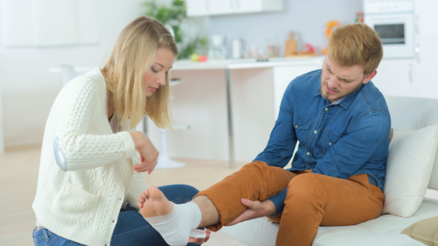
  • Protection: after an acute injury, an athlete should remove themselves from play and protect the injured area from additional damage by applying a bandage, elastic wrap, sling, or splint, or even tape may do the job; if the injury is to any part of the leg, avoid bearing weight on that side and consider using crutches
  • Rest: it’s important to take a break from your respective sport in order to allow your body to heal; the amount of rest needed depends on the type and severity of the injury, but if participation leads to pain, it’s wise to continue resting; keep in mind that this doesn’t necessarily mean you need to avoid all activities, but you should avoid those that can further aggravate your injury
  • Ice: cold treatment, or cryotherapy, is one of the easiest but most effective ways to reduce pain and other symptoms immediately after an injury; applying ice slows down blood flow to the injured area and in effect reduces inflammation and swelling, as well as muscle spasms; ice should be used as soon as possible after the injury and applied for 15-20 minutes every 1-2 hours for the first few days
  • Compression: pressure also helps to reduce inflammation and swelling, while also providing minimal support to the injured area; in most cases a simple elastic bandage will be sufficient, which should be applied snugly but not too tight, and directly to the skin by starting a few inches below the injury and wrapping in a spiral to a few inches above the injured area
    • There are also devices available designed specifically for compression that provide an even amount of pressure around an injured area for added benefits; some of these devices combine compression with cold therapy, which temporarily reduces blood flow and allows the cold to penetrate through the skin and deep into the muscles, ligaments, and bones
  • Elevation: by elevating the injured body part, you will be draining the pooling of fluids away from that area, which reduces swelling, inflammation and pain; this can be accomplished by positioning the injured area above the level of the heart for as much time as possible for at least the first 48 hours after the injury

Other home remedies to try right after an injury

The PRICE protocol can be supplemented with a number of other remedies that you can usually perform on your own at home, including:

  • Heat therapy: applying heat (thermotherapy) increases the flow of blood to the injured area, which in turn stimulates the body’s natural healing mechanisms, while also reducing pain and stiffness; thermotherapy can be applied at similar intervals to cryotherapy (15-20 minutes every 1-2 hours), but should not be used until after the initial inflammation from the injury has begun to die down (usually a few days)
  • Contrast therapy: switching back and forth between cryotherapy and thermotherapy alternately opens and constricts the blood vessels and increases blood flow to an injured area without causing additional fluids to accumulate
  • Pain medications: acetaminophen (Tylenol) may be best for the first day, as it reduces pain without increasing bleeding; after the first day or two, aspirin or non-steroidal anti-inflammatory drugs (NSAIDs) like ibuprofen (Advil) and naproxen (Aleve) will help to alleviate both inflammation and pain
  • Massage: lightly massaging the injured area may help prevent the build-up of fluid that leads to swelling

While these simple remedies are best for mild injuries, more severe injuries require care from a trained professional. In these cases, it’s best to see a physical therapist, who will perform a thorough evaluation of your condition and create a personalized and comprehensive treatment program based on your goals and abilities.