Physical therapy can help avoid the expenses of surgery

Undergoing physical therapy can help avoid the expenses and potential risks associated with surgery

If you experience an injury or happen to be in pain for a long period of time, relief may eventually become your number one priority. There are many treatment options available to address these types of issues, and what’s right for you depends on several important individual factors. But in nearly every case, one thing remains true: trying physical therapy first is a smart move that will usually help you avoid other unnecessary tests and treatments.

Physical therapy is not a magical cure-all that will immediately fix any physical problem you have, but it does have an incredibly wide range of applications, and it can be used to treat patients of any age and activity level. Every physical therapy treatment program is individually tailored with each patient’s abilities and goals in mind, and by carefully guiding them with exercises and movement-based techniques, significant improvements can usually be expected after treatment is completed.

Another route that some patients may follow is to have surgery to address their injury or painful condition. Surgery has great value that can often lead to positive outcomes, and it may be necessary in certain situations, but it does come with some potential downsides as well. The cost of most surgeries is usually quite high, recovery time can be extensive, and there are also some risks involved with the procedure itself. Physical therapy, on the other hand, is universally regarded as an affordable, safe intervention with minimal to no associated risks. In addition, physical therapy can lead to similar—or sometimes better—outcomes compared to surgery, and in some cases it can actually help patients avoid the need for surgery altogether when they access it first. Below are a few studies that highlight some of the advantages of physical therapy over surgery:

How does surgery compare to sham surgery or physiotherapy as a treatment for tendinopathy? A systematic review of randomised trials (2019)

  • How the study was conducted: this was a systematic review, which collects and analyzes the findings of several studies on the same topic; in this case, 12 studies were evaluated that compared surgery and physical therapy for patients with various types of tendinopathy (an injury involving a tendon)
  • What the results showed: physical therapy was found to be just as effective as surgery in the midterm and long term for reducing patients’ pain and improving their function and flexibility

Does Anterior Cruciate Ligament Reconstruction Improve Functional and Radiographic Outcomes Over Nonoperative Management 5 Years After Injury? (2018)

  • How the study was conducted: 105 patients with a torn anterior cruciate ligament (ACL) underwent either physical therapy or surgery to treat their injury; five years later, they were evaluated with an MRI and several other tests
  • What the results showed: after five years, there were only minimal differences between patients treated surgically compared to those who had physical therapy

Arthroscopic Surgery or Physical Therapy for Patients With Femoroacetabular Impingement Syndrome: A Randomized Controlled Trial With 2-Year Follow-up. (2018)

  • How the study was conducted: 80 patients with a hip condition called femoroacetabular impingement syndrome were randomly assigned to undergo either surgery or physical therapy; then for two years, these patients were evaluated regularly to compare their outcomes
  • What the results showed: all patients improved significantly, there were no significant differences between the two groups after two years, and no clear advantages of surgery were found

Association of Early Outpatient Rehabilitation With Health Service Utilization in Managing Medicare Beneficiaries With Nontraumatic Knee Pain: Retrospective Cohort Study (2017)

  • How the study was conducted: the healthcare usage of patients with knee pain was evaluated over 12 months and categorized according to if and when they underwent physical therapy
  • What the results showed: patients who had early physical therapy were found to be 42% less likely to undergo surgery than those who did not

These are just a few examples in the research that show why physical therapy is a better option than surgery for any injury or painful condition you might be dealing with. We’d also like to remind you that now is a great time of year to see a physical therapist for those trying to get the most out of their healthcare plan. If you haven’t done so already, review your health insurance policy and check on your benefit status now. If you’ve already met your deductible or out-of-pocket maximum for 2019, you will likely have a lower co-pay or no co-pay at all for the rest of the year, before your deductible renews on January 1, 2020.

For patients who repeatedly sprain their ankles hands-on therapy lead

Many patients who sprain their ankle once have long-term issues

Ankle sprains are the most common injury in athletes and those who are physically active. In addition to the initial pain and disability that these injuries cause, up to 41% of patients who sprain their ankle once will go on to develop a condition called chronic ankle instability (CAI). Individuals with CAI tend to experience continual pain and soreness, and a sensation that the ankle is giving out. As a result, they are more likely to sprain their ankle again, which can lead to serious long-term disability. Exercise therapy is one of the more commonly used treatments for patients with CAI, with exercises that strengthen muscles and improve balance and proprioception (the sense of knowing where one’s body is in space) being most effective. Another popular treatment is called manual therapy, in which a physical therapist moves and manipulates certain joints in specific ways to improve their mobility and alleviate symptoms. In some cases, manual therapy will target the nerves as well as the joints and soft tissue of a region. Although research has shown both exercise and manual therapy to be effective for patients with CAI, no studies have evaluated manual therapy that focuses on the nerves of the ankle. For this reason, a powerful study called a randomized-controlled trial (RCT) was conducted to determine if manual therapy can provide any additional benefits to an exercise program for CAI patients.

Patients are randomly divided into two groups

Patients with CAI were invited to participate in this study and then screened with specific criteria to determine if they were eligible. This process led to 56 patients being included in the study and then randomly assigned to one of two groups: experimental group I or experimental group II. Under this design, all patients participated in an exercise program that took place for four weeks, but the experimental group II also received a course of manual therapy. The exercise program consisted of six exercises that were repeated during two sessions per week and got progressively more difficult each week. These exercises—which a physical therapist supervised—focused on strengthening the muscles surrounding the foot and ankle and improving each patient’s proprioception, which is closely related to balance. Manual therapy consisted of several techniques applied to the joints and nerves of the ankles, which lasted for 20-30 seconds each with two minutes of resting in between. All patients were assessed before each treatment program, immediately afterwards, and then one week later for a number of outcomes, including pain, strength, ankle instability, and flexibility.

Greater benefits are achieved when exercise is combined with manual therapy

One week after treatment ended, it was found that patients in both groups improved in all of the outcomes that were measured; however, patients in experimental group II experienced greater improvements than those in experimental group I. This was found to be the case in most of the measurements taken, but was particularly true for ankle instability and strength, both of which had scores that were considerably higher in the group that underwent manual therapy. These results suggest that although exercise therapy is capable of producing benefits in patients with CAI, adding manual therapy to this exercise program leads to even better overall outcomes. Individuals who suffer from CAI are therefore encouraged to see a physical therapist if this step has not already been taken. Physical therapists are movement experts that can diagnose your condition and provide you with a comprehensive, personalized treatment program involving these components that will set you up for a positive outcome.

-As reported in the December ’16 issue of Manual Therapy

For patients who repeatedly sprain their ankles hands-on PT can help

Many patients who sprain their ankle once have long-term issues

Ankle sprains are the most common injury in athletes and those who are physically active. In addition to the initial pain and disability that these injuries cause, up to 41% of patients who sprain their ankle once will go on to develop a condition called chronic ankle instability (CAI). Individuals with CAI tend to experience continual pain and soreness, and a sensation that the ankle is giving out. As a result, they are more likely to sprain their ankle again, which can lead to serious long-term disability. Exercise therapy is one of the more commonly used treatments for patients with CAI, with exercises that strengthen muscles and improve balance and proprioception (the sense of knowing where one’s body is in space) being most effective. Another popular treatment is called manual therapy, in which a physical therapist moves and manipulates certain joints in specific ways to improve their mobility and alleviate symptoms. In some cases, manual therapy will target the nerves as well as the joints and soft tissue of a region. Although research has shown both exercise and manual therapy to be effective for patients with CAI, no studies have evaluated manual therapy that focuses on the nerves of the ankle. For this reason, a powerful study called a randomized-controlled trial (RCT) was conducted to determine if manual therapy can provide any additional benefits to an exercise program for CAI patients.

Patients are randomly divided into two groups

Patients with CAI were invited to participate in this study and then screened with specific criteria to determine if they were eligible. This process led to 56 patients being included in the study and then randomly assigned to one of two groups: experimental group I or experimental group II. Under this design, all patients participated in an exercise program that took place for four weeks, but the experimental group II also received a course of manual therapy. The exercise program consisted of six exercises that were repeated during two sessions per week and got progressively more difficult each week. These exercises—which a physical therapist supervised—focused on strengthening the muscles surrounding the foot and ankle and improving each patient’s proprioception, which is closely related to balance. Manual therapy consisted of several techniques applied to the joints and nerves of the ankles, which lasted for 20-30 seconds each with two minutes of resting in between. All patients were assessed before each treatment program, immediately afterwards, and then one week later for a number of outcomes, including pain, strength, ankle instability, and flexibility.

Greater benefits are achieved when exercise is combined with manual therapy

One week after treatment ended, it was found that patients in both groups improved in all of the outcomes that were measured; however, patients in experimental group II experienced greater improvements than those in experimental group I. This was found to be the case in most of the measurements taken, but was particularly true for ankle instability and strength, both of which had scores that were considerably higher in the group that underwent manual therapy. These results suggest that although exercise therapy is capable of producing benefits in patients with CAI, adding manual therapy to this exercise program leads to even better overall outcomes. Individuals who suffer from CAI are therefore encouraged to see a physical therapist if this step has not already been taken. Physical therapists are movement experts that can diagnose your condition and provide you with a comprehensive, personalized treatment program involving these components that will set you up for a positive outcome.

-As reported in the December ’16 issue of Manual Therapy

For ankle/foot pain, rest, massage, and footwear adjustments are best

Try to think about all the movements that are needed to get you through a typical day. Chances are, regardless of what type of work you do or what your day consists of, you’ll notice that you probably use your feet and ankles at least a fair amount. This is even more true for active individuals, as most forms of physical activity will require some walking, running, and possibly even jumping to complete.

It’s easy to overlook the significant amount of work that the feet and ankles are doing to keep you stable and allow you to get around, but this can all change when pain enters the picture. Dealing with a sore ankle or nagging heel pain that hits you the first thing in the morning will likely alter your entire perspective and force you to make some adjustments to your daily routine to make moving easier.

Foot and ankle pain are quite common and can result from a number of different injuries, but the end result is usually a limited ability to live out your day as you’d like. For this reason, when it does strike, your next question is probably related to making it dissipate as quickly as possible. Fortunately, there are a number of steps you can take on your own to address an ankle or foot injury and get you back to full strength quickly. We recommend the following:

  • Adhere to the RICE protocol: after traumatic injuries—especially ankle sprains—your first step should always be to respond with the RICE protocol within the first 24-72 hours; doing so will relieve painful symptoms and reduce your risk for further damage to the area during this time
    • Rest: take some time to rest and recuperate immediately after the injury and avoid any activities that can aggravate your pain; this can range from a few days to a week or more, depending on the injury; for severe ankle sprains, crutches may be needed to help you avoid putting pressure on the ankle
    • Ice: in the first few days after a traumatic injury, ice is your friend, as it will slow down blood flow and reduce inflammation, swelling, and muscle spasms; start using it right after the injury and apply it for 15-20 minutes every 1-2 hours during this time
    • Compression: after an ankle sprain, wrap an elastic bandage snugly around your ankle to help reduce inflammation and swelling; for severe ankle sprains, an ankle brace may be needed, which adds further protection from future injury
    • Elevation: within the first 48 hours after an injury, elevate your foot above your head for as much time as you can manage to drain the pooling of fluids away from the region and reduce swelling, inflammation, and pain
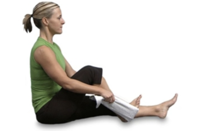
  • Massage: if you’re experiencing pain in one particular region, massage can help by improving circulation and reducing your soreness; for plantar fasciitis—for example—rub and knead the bottom of your foot near the heel with ample pressure; using a frozen water bottle to massage your foot is doubly effective because it also applies cold therapy to the area
  • Add shoe inserts: also known as insoles, arch supports, or orthotics, these devices can provide your foot with extra cushioning and added support; using an orthotic is particularly effective for individuals with plantar fasciitis due to flat feet
  • Replace your shoes: sometimes orthotics won’t be enough; if the soles of your shoes are worn out and not providing you with enough support, or if you notice that your feet are in pain after every time you use your shoes, it may be time to purchase a new pair; when buying athletic shoes, it’s best to go to a running store that has the capability of analyzing your gait to ensure you’re using the right pair for your foot type
  • Consider pain medications: depending on the severity of your issue, you may experience some relief with over-the-counter pain medications; non-steroidal anti-inflammatory drugs (NSAIDs) like aspirin, ibuprofen (Advil), and naproxen (Aleve) will reduce inflammation in addition to pain
  • Wear a night splint: for severe plantar fasciitis, a night splint may help by keeping your foot in a locked position overnight; this can prevent you from pointing your foot, and in effect, alleviate pain

Treat your feet right and stay conditioned to avoid injury

For most of us our feet provide the freedom to get from point A to B with minimal effort. But even though they provide this incredible ability, it’s easy to take the feet for granted, and perhaps we often ignore these wonderful appendages.

It becomes more difficult to disregard the feet, though, when something goes wrong with them. This is particularly the case when a foot or ankle injury prevents one from walking or running normally, leading to a redirection of the attention downwards.

Injuries to the feet and ankle actually rank among some of the most common in the entire body because they handle so much weight and are used so frequently. Ankle sprains are by far the most prevalent injury to this region—especially in active individuals—while others include plantar fasciitis, Achilles tendinitis, stress fractures, and growth plate injuries (in children). Unfortunately, there’s no magic formula that will prevent all foot and ankle injuries from occurring, but there are a number of steps you can take to significantly reduce your personal risk for experiencing one. We recommend the following:

  • Warm up and stretch: regardless of what activity you’re about to partake in, it’s always important to get the blood flowing with a warm-up and some stretching before physically exerting yourself; dynamic stretches that mimic the sport or exercise you’re participating in are best for reducing injury risk
  • Build up your strength: strengthening the muscles of the feet and lower leg is extremely important for injury prevention, as it will provide a more sturdy foundation for the rest of the body; strong muscles in this area will also improve balance, which can further reduce injury risk
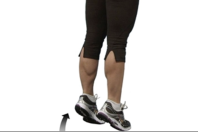
    • Calf raises: stand with your feet shoulder-width apart, keeping the knees straight; raise the heels off the floor as high as you can, then return to the floor and repeat; to progress the exercise, stand with your toes on a step
    • Toe splay: sit in a straight-backed chair with your feet gently resting on the floor; spread your toes apart as far as possible without straining them and hold the position for 5 seconds, then repeat this motion 10 times
    • Resisted ankle inversion: sit on the floor with your legs outstretched and a band wrapped around the foot you want to work; tie the other end around something sturdy to the outside of the leg; keep the lower leg still and try to point the toes across to the other leg
    • Resisted ankle eversion: for eversion, move the attachment of the resistance band so it’s on the other side of the body, then turn the foot out and try to point your toes away from the other foot
  • Wear the right shoes: try to always use shoes that are appropriate for the activity; your shoes should provide support and comfort while being able to withstand the physical demands of the sport they’re being used for; it’s also a good idea to avoid wearing high heels regularly and to be careful about your sandal selection
  • Steer clear of the wrong surfaces: running or training on uneven surfaces can increase the likelihood of an injury, so it’s best to avoid these as much as possible
  • Train in moderation: many injuries result from overdoing it or increasing physical activity levels too aggressively; this is true for everyone, but especially those who have not been active in a long time and those starting a new sport or activity; try to advance your regimen gradually to avoid these types of injuries
  • Listen to your body: if you do notice pain in your foot or ankle, it’s probably a sign from your body that you’re overdoing it; learn the difference between typical muscle soreness (which often develops after working out) and lingering pain (which could be a sign of a more serious issue), and seek out help when the pain doesn’t subside

Foot & ankle injuries are a common problem for active individuals

foot & ankle injuries

Over time, exercise and physical activity tend to strain certain areas of the body to the point of pain, and this distribution is not equally divided. While the region that’s affected has much to do with the activity being performed, there are some spots that just seem to carry the brunt of the load no matter what.

The feet and ankles clearly fit this bill, as they are some of the most common locations for injury throughout the body. Problems arising here can occur in anyone, but those who keep an active lifestyle are far more likely to be affected than others. This is apparent across the age spectrum, as foot and ankle injuries are seen frequently in children and then remain prevalent into adulthood, with some people experiencing the same issues in the long term.

Understanding a few of the most common foot and ankle injuries at different ages is helpful, as it can prepare you to identify the signs of a problem and learn how to respond appropriately.

Developing bodies and high levels of activity contribute to risk in children

Children and adolescents often tend to have a seemingly never-ending supply of energy, some of which is spent on sports and exercise, among other things. The high participation levels in sports and the generally active habits of kids are the primary reasons foot and ankle injuries occur so frequently. But in addition, children’s bodies are still growing and have not yet fully developed, which increases their risk for injury even further. Below are some of the injuries that are most likely to be seen in this age group:

  • Sever’s disease: this overuse injury results from inflammation of the growth plate in the heel, which is an area of growing tissue near the ends of bones in children; these injuries are caused by repetitive stress to the heel and are most likely to occur during growth spurts, when a child’s heel bone grows faster than the muscles, tendons, and ligaments; symptoms include pain and tenderness underneath the heel
  • Ankle sprain: ankle sprains occur when the ligaments surrounding the ankle—which connect bones to bones—are stretched beyond their limit in a forceful motion; these are the most common injuries in all of sports, and they typically lead to pain, swelling, and an inability to put pressure on the ankle
  • Growth plate fracture: since growth plates are weaker than tendons and ligaments, incidents that would normally lead to an ankle sprain in older individuals could actually cause a growth plate fracture instead in children and adolescents; in some cases, both injuries occur at the same time
  • Stress fractures: these are small cracks or severe bruising within a bone that are caused by repetitive stress or force to the foot; they are particularly common in older adolescents who are extremely active in sports like soccer and gymnastics

Some problems remain common, while other new ones arise later in life

Foot and ankle injuries remain problematic later in life, too. Growth plate injuries are no longer a possibility since the plates are fully developed by adulthood, but a number of other issues become more likely in older age and repeated trauma to these regions. Among those are the following:

  • Ankle sprain: unsurprisingly, ankle sprains are still the most common sports-related injury in adults, just like children; in fact, about half of all ankle sprains are related to physical activity, and about 25,000 people sprain their ankle everyday; since growth plates are not present, twisting motions to the ankle typically result in a sprain or fracture of the ankle
  • Plantar fasciitis: generally considered to be the most common cause of heel pain in adults, this condition results from inflammation of the plantar fascia, a thick band of tissue that connects the heel to the toes; when this tissue is overstrained from repeated activity—like running—it becomes inflamed, which leads to a stabbing pain near the heel that’s most noticeable upon waking up
  • Achilles tendinitis: another overuse injury related to inflammation of the Achilles tendon, which connects the calf muscle to the back of the heel; it’s most common in runners who do lots of speed training, uphill running, or rapidly increase their training intensity or duration, and it leads to heel pain that usually comes on gradually as a mild ache in the back of the leg or above the heel
  • Stress fractures: these injuries also remain common in adults and are most frequently seen when the bones and their supporting muscles don’t have time to heal between exercise sessions, or when a person changes their usual exercise regimen with a sudden increase of activity or a change in workout surface
  • Turf toe: this is a sprain of the ligaments surrounding the big toe when it’s bent back too far (hyperextended), which is common in football players; it can occur from a sudden, forceful movement or repeated hyperextensions over a period of time, and leads to pain, swelling, and limited movement of the big toe

PT leads to improvements for patients with common jaw disorder

More research is needed that evaluates how effective these treatments are

Temporomandibular disorder (TMD) is a term used to describe a set of common conditions that affect the temporomandibular joint (TMJ). The TMJ connects the jaw to the skull and allows it to move up and down and from side to side. Typical symptoms of TMDs include pain (especially while chewing), difficulty opening the mouth, the jaw getting stuck, and a clicking or popping sound, all of which can have a negative impact on patients’ lives. There are a number of available treatments for TMDs, two of which are physical therapy and occlusal splints. Physical therapy usually consists of a variety of interventions that are all intended to reduce pain and improve strength, mobility, coordination, posture, and flexibility. An occlusal splint is a specific type of mouth guard that prevents clenching the jaw and protects the teeth from other harmful habits. While both of these treatments are commonly used for treating TMDs, there is no research that compares the two of them. For this reason, a powerful study called a randomized-controlled trial (RCT) was conducted to determine if physical therapy or wearing a splint is more effective for patients with a TMD.

Both interventions last for six weeks

To conduct the study, researchers recruited patients who were diagnosed with a TMD and screened them using specific criteria to determine if they could participate. From this search, 112 individuals were accepted to the study and randomly assigned to either the physical therapy group or the splint group. The physical therapy treatment program took place during three 15-minute sessions per week for six weeks. During these sessions, a physical therapist led patients through a series of exercises and stretches to relax the jaw. In the relaxed jaw position, for example, patients were told to place their tongue behind their upper front teeth and allow the teeth to come apart in order to relax the jaw muscles. In the splinting group, patients were given an occlusive splint and told to wear it every day for the next six weeks as prescribed by a dentist. Measurements of pain levels and jaw flexibility were taken for all patients at the start of the study and then again at the end of the interventions six weeks later.

Patients with a TMD should see a physical therapist to receive similar treatments

When patients were assessed at the end of the study, it was found that those who followed a course of physical therapy experienced significantly greater improvements compared to those who wore a splint. This was found to be the case for both pain levels and flexibility of the jaw, the two outcomes that were measured. These results suggest that physical therapy provides superior outcomes for patients with a TMD than wearing an occlusal splint. Physical therapy has also been found to be a safe, relatively easy, and inexpensive intervention, which makes it even more attractive. For these reasons, if you currently have a TMD and are bothered by pain, it’s recommended that you see a physical therapist. Doing so will get you on the right track and provide you with the treatment needed to manage your symptoms and regain the ability to move your jaw without pain.

-As reported in the September ’18 issue of The Journal of Physical Therapy Science

You can alleviate your jaw pain with some simple home remedies

Whether you realize it or not, you use your temporomandibular joint (TMJ) pretty often. This joint connects your lower jaw to the rest of the skull right in front of your ears, and you can actually feel it if you touch right below your temples and open your mouth. The TMJ allows the jaw to move up and down and from side to side, which is why it’s used with practically every movement that involves your mouth. So when you speak, swallow, or bite down on something, you have both of your TMJs to thank.

Unfortunately, the TMJ also has a reputation of being a common location for pain. Temporomandibular disorder (TMD) is a general term used to describe any condition that affects the TMJ. The common trait of all TMDs is pain and possibly inflammation in the muscles of the jaw and surrounding area. This pain can spread to the cheek, ear, or temple, and it often causes difficulty performing any tasks that require opening or closing the mouth. As with many other parts of the body, you might not become acquainted with your TMJ until something goes wrong with it, as is the case with these conditions.

Approximately 5-12% of the population is affected by a TMD, with the majority of these individuals dealing with acute pain—meaning it developed recently. If you count yourself as one of the millions of Americans dealing with a TMD right now, you may be curious if there are any simple solutions out there that will help to alleviate your symptoms. The good news is yes, there are. Most cases of acute pain from a TMD can be effectively treated with some home remedies that you can perform on your own. We recommend the following tips for a recent onset of pain or other symptoms to the TMJ:

  • Heat therapy: applying heat with a moist warm towel or dry heating pad can reduce jaw pain and stiffness by increasing the flow of blood to the area; it’s recommended that you use a warm compress and place it on the jaw and temples for about 10-15 minutes, about twice a day; heat therapy is particularly effective if you have a dull, steady, aching pain
  • Ice therapy: if the heat does not lead to any notable improvements after two days, try applying ice instead; the application of ice will numb the nerves and dull the pain by slowing down the flow of blood to the area; apply an icepack wrapped in a towel to the area for about 15-20 minutes a few times a day
  • Massage: massaging the areas around your jaws can also improve blood flow and provide relief; this can be accomplished by opening your mouth and locating the muscles next to your ears by the TMJ; put your fingers on any area that’s sore and apply gentle pressure in a circular motion; you can also massage the muscles on the sides of your neck if there is tension in that region as well
  • Jaw exercises: regularly moving the jaw in a specific and systematic manner may be helpful for further reducing TMD symptoms; a selection of exercises is below
    • Relaxed jaw exercise: rest your tongue gently on the top of your mouth behind your upper front teeth; allow your teeth to come apart while relaxing your jaw muscles
    • Chin tucks: with your shoulders back and chest up, pull your chin straight back, creating a “double chin;” hold for three seconds and repeat 10 times
    • Resisted opening of the mouth: place your thumb under your chin; open your mouth slowly, pushing gently against your chin for resistance; hold for three to six seconds, and then close your mouth slowly
    • Resisted closing of the mouth: squeeze your chin with your index and thumb with one hand; close your mouth as you place gently pressure on your chin; this will help strengthen your muscles that help you chew
  • Pain-relieving medications: non-steroidal anti-inflammatory drugs (NSAIDs) like ibuprofen and other over-the-counter pain medications may help to reduce inflammation and alleviate pain in some individuals
  • Splint or night guard: these devices fit over your upper and lower teeth to prevent these rows from touching one another; in effect, this will lessen the impact of clenching or grinding the teeth and correct your bite by putting your teeth in a better position

Avoid a jaw problem by chewing properly and practicing good posture

There are over 200 bones in the human body, and the head and face account for 22 of these. Of the face and head bones, the lower jawbone—or mandible—is the only one that can move. The mandible is connected to the temporal bone of the skull at two points just in front of each ear through an important joint called the temporomandibular joint. This joint gets lots of attention not only because it allows us to move our jaw in order to talk and chew food, but also because it’s a common location for pain.

A temporomandibular disorder (TMD) is essentially any condition in which the temporomandibular joint is not functioning properly. These types of issues are quite common, as they affect millions of Americans every year and can strike at any age. Symptoms vary from patient to patient, but in most cases they lead to pain and discomfort in the jaw and surrounding facial muscles which can make it difficult to perform any actions that require opening or closing the mouth.

It’s not entirely clear what causes TMDs, but several factors may be responsible, including injury to the jaw, repeated clenching or grinding of the teeth, high stress levels, muscle spasms, and arthritis. This uncertainty, combined with the fact that we need to use our jaws constantly, might lead you to believe that there’s not much that can be done to avoid a TMD. Fortunately this is not the case, as there are several steps you can take to reduce your risk for TMDs. We recommend the following strategies:

  • Be more careful with what and how you chew
    • Avoid chewing gum
    • Chew with both sides of your mouth
    • Avoid eating too many hard or crunchy foods
    • Take smaller bites of food
    • Don’t bite on hard objects like pens or pencils
    • Avoid biting your nails
  • Practice good posture
    • Keep your head balanced and not hunched forward, your shoulders straight, and torso in alignment with your head and shoulders (with good posture, a straight line can be drawn from your ears to your shoulders)
    • Avoid cradling your phone between your neck and shoulders
    • Try not to regularly carry a heavy purse or backpack on one shoulder
    • Consider using ergonomically-designed products for your office setup
  • If you grind or clench your teeth, try to reduce or stop this habit
  • Try to keep your tongue at the roof of your mouth and avoid letting your teeth touch; your teeth should be kept at least a few millimeters apart unless you’re chewing, and even then they should not be making much contact with one another
  • Sleep on your back or side and avoid sleeping on your stomach, which can strain your jaw; make sure your pillow provides enough support for your head and neck
  • If your stress levels are high, explore options to reduce your stress, including meditation, yoga, mindfulness practices, and cognitive behavioral therapy
  • Don’t rest your chin in your hands
  • Breathe through your nose and keep your lips together

Jaw disorders can cause problems for similar reasons at different ages

Pain can develop just about anywhere in your body, and regardless of where it occurs, can prove to be an annoying problem. The jaw is one region that doesn’t get discussed as often as more common injury sites like the knees or the neck, but disorders of the jaw are actually more prevalent than you may think. These types of issues—which are called temporomandibular disorders—affect millions of Americans every year and typically cause nagging symptoms like jaw pain and headaches. Some people are more likely than others to have a problem with their jaw, but they can occur in anyone and at any age.

The temporomandibular joint (TMJ) is a hinge joint that connects the part of the skull directly in front of the ears (temporal bone) to the lower jaw (mandible). It allows you to move your jaw up and down and from one side to the other, which is necessary for talking and chewing. Temporomandibular disorder, or TMD, is a general term used to describe a variety of conditions that cause pain and dysfunction in the jaw, jaw joint, and surrounding facial muscles that control chewing and jaw movement.

The definite cause of TMDs is still unclear, but some theories suggest that they may be due to injury in that region, grinding, or clenching teeth, osteoarthritis, or stress. Symptoms vary from patient to patient, but some of the more common signs of a TMD include the following:

  • Jaw pain or tenderness, which can be on one or both sides of the jaw
  • Aching pain in/around your ear or in your face
  • Difficulty opening/closing the mouth or chewing
  • Popping, clicking, or locking of the jaw
  • Headaches
  • Ear aches or ringing in the ear

TMDs can occur as early as infancy, but are not frequently seen in this age group and are more likely to develop somewhat later in life. When a newborn or young child is diagnosed with a TMD, it’s typically related to a deformity that was present at birth. But as children age, the likelihood of experiencing a TMD increases, particularly around the teenage years. Girls are more likely than boys to have an issue with their jaw.

It’s difficult to define exactly what causes a TMD in children and adolescents, but most experts believe that overexerting the jaw is at least partially responsible. Overexertion can occur from high levels of stress or anxiety, repeatedly clenching the jaw or grinding teeth—which is called bruxism—or from a traumatic injury such as one sustained in sports. High stress levels can actually make kids more likely to tighten their jaws, and over time, these behaviors will change the alignment of one’s bite and affect the muscles used for chewing. Regularly using a forward head posture with slouched shoulders and a rounded upper back may also contribute to the development of TMDs, since this type of posture can affect how the jaw closes.

The risk for TMDs continues to increase to a certain point, as the majority of cases are seen between the ages of 20-40. About 15% of adults currently suffer from a TMD, and once again, women are at least twice as likely to get them than men. As with younger populations, a highly stressful lifestyle, injuries to the jaw, and frequent jaw clenching will all remain factors that could play a role in the development of a jaw problem. Unregulated stress and a long-term habit of jaw clenching will likely increase this risk even more on account of the repeated stress placed on the jaw over time.

But there are also some risk factors that are unique to adults and may also increase the risk for developing a TMD. Arthritis—which includes rheumatoid arthritis, osteoarthritis, and gouty arthritis—is more likely to occur later in life. These types of arthritis can affect various joints of the body, and if one of the TMJs is involved, the risk for experiencing a TMD may be higher. Muscle spasms caused by other medical conditions or injuries may, as well as changes in the structure of teeth from dental procedures or the wearing down of teeth may also become more likely in older age. Both of these factors can also play a part in the development of a TMD.