October is a special time for our profession that weâd like you to be a part of. It is National Physical Therapy Month, which is an annual opportunity for physical therapists and physical therapist assistants around the country to campaign and speak out about our profession. The goal of the monthâlong campaign is to increase awareness about the important role physical therapy can play in peopleâs lives by reducing pain, improving mobility, and encouraging a healthy lifestyle.
National Physical Therapy Month was first established in 1981 by the American Physical Therapy Association (APTA), when the organization wanted to show the public how physical therapy can âtransform society by optimizing movement to improve the human experience.â In doing our part to work towards these goals, over the next few posts we will be providing an overview of what physical therapists do and the wide range of conditions they treat throughout the entire body.
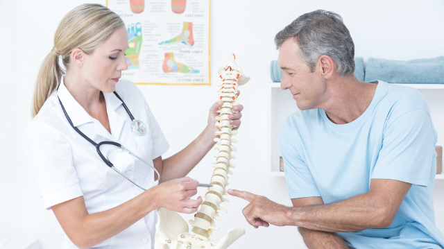
Understanding the many conditions that involve the neck or back
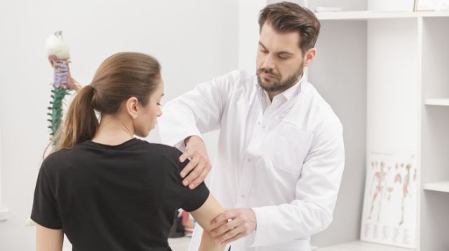
Physical therapists are experts in how the body moves. When a patient comes to us with an injury or painful condition, we first perform a thorough evaluation to identify the source of the problem and then create a customâtailored program that targets the patientâs impairments and limitations. While the elements of each program will vary depending on the specific condition and the patientâs unique needs, abilities, and goals, physical therapists always provide a highly personalized approach to care and pay close attention to each patientâs response, regardless of what theyâre being treated for or the location of pain.
In our first post of this series, weâre going to explore which conditions of the spine physical therapists most frequently treat.
The spine is one of the most common locations in the body where pain can arise. Up to 50% of adults deal with neck pain each year, and up to 70% will encounter it at least once in their lifetime. The figures on back pain are even higher, as about 80% of Americans will experience an episode of low back pain at some point in their lives, making it the most common site for pain in the body. Many of the ailments that produce pain in the neck can also develop in the back, and vice versa. Letâs take a look at some of the most prevalent spineârelated conditions:
- Sprain: occurs when a ligament in the spine is pushed beyond its limits, which can cause it to be damaged or torn; typically leads to pain, discomfort, reduced range of motion, and possibly muscle cramping or spasms
- Strain: involves a tendon or muscle that supports the spine being twisted, pulled, or torn; as with sprains, neck and back pains usually lead to pain, discomfort, reduced range of motion, and possibly muscle cramping or spasms
- Both sprains and strains can occur either from a single incident or due to repetitive stress over time, and these injuries are responsible for most cases of neck and back pain, particularly in younger patients
Herniated disc: a condition in which the softer jellyâlike substance of a disc in the spine pushes out through a crack in the tough exterior ring; a âbulging discâ means that the inner layer has protruded outwards, but the outer layer remains intact; a herniated disc is most likely to occur in the lower back, but they are also seen in the neck; common symptoms include arm or leg pain, numbness or tingling, and weakness
Spinal stenosis: a condition in which the spinal canalâthe space around the spinal cord filled with a fluid that bathes the nerves and nerve roots of the spineânarrows over time, which puts pressure on the spinal cord and spinal nerve roots; spinal stenosis is most common in the lower back and the neck and is typically only seen in older adults since itâs caused by ageârelated changes
Joint dysfunction: a term used to describe when any of the joints of the spineâincluding the facet joints or sacroiliac jointâare either moving too much or too little; this can lead to pain and other symptoms in the hips, pelvis, and lower back
Degenerative disc disease: an ageârelated disorder in which one or more of the intervertebral discs deteriorates or breaks down, which can lead to a herniated disc or other related issues; degenerative disc disease is another one of the most common causes of low back and neck pain
Osteoarthritis: involves the breakdown of protective cartilage that surrounds the ends of joints and discs in the spine; osteoarthritis can occur anywhere in the spine, and has been referred to as the most common cause of low back pain in people over the age of 50; patients typically experience pain and stiffness, as well as weakness or numbness in some cases
Spondylosis: a general term used to describe any pain related to ageârelated changes in the spine; can occur in the neck or back
Evidence supports the effectiveness of physical therapy for neck and back pain
Physical therapists utilize a variety of interventions to address neck and back pain, including stretching and strengthening exercises, manual (handsâon) therapy techniques, painârelieving modalities, functional training, education, and guidance on how to avoid further aggravation of pain. After adhering to these treatment recommendations, patients will eventually notice a marked reduction in their pain levels while gradually regaining the ability to move and function similarly to before the onset of their pain.
Research has shown that physical therapy can lead to a multitude of benefits for patients with back or neck pain, including less pain and disability, lower overall treatment costs, and a lower chance of needing additional treatments while avoiding both surgery and opioids.
In our next post, weâll review conditions that affect the shoulder, elbow, and wrist.