Every 34 seconds, one American dies from cardiovascular disease. This makes cardiovascular disease—or heart disease—the leading cause of death in the U.S. for men and women and for most ethnic and racial groups, with about 700,000 people dying from it each year.
The magnitude of this problem cannot be overstated, but it’s equally important to note that many of these deaths can be prevented through certain lifestyle changes.
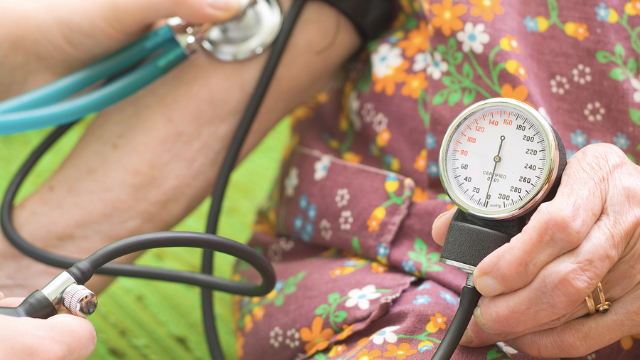
One of the leading factors risk factors for heart disease is high blood pressure (or hypertension). Often referred to as “the silent killer,” hypertension is a disease in which blood flows through arteries at a pressure that is higher than normal. Blood pressure is measured with two numbers, and normal pressure is 120/80 or lower, while stage 1 high blood pressure is any reading 130/80 or higher and stage 2 is 140/90 or higher. High blood pressure may not cause any symptoms—which explains its nickname—but it significantly increases one’s risk for heart disease, heart failure, stroke, and other cardiovascular complications.
The risk for hypertension increases with age, and it’s more likely to occur in men before the age of 64 but more common in women after the age of 65. It is also more common in Black individuals—often developing at an earlier age compared to White individuals—and in those who have a parent or sibling with the condition. These are all considered “nonmodifiable” risk factors for hypertension because they are out of each patient’s control; however, there are also many modifiable risk factors for hypertension that every individual has the power to address.
Some of the most dangerous modifiable risk factors for hypertension are being overweight or obese, not exercising, eating a poor diet, having high levels of stress, and smoking. We’re going to focus on diet and exercise since addressing these factors is not only helpful for reducing one’s risk for hypertension, but also essential for good overall health.
Diet
Reducing inflammation and oxidative stress are keys to mitigating the risk for hypertension, which is primarily accomplished through a well‐balanced diet and regular exercise. Knowing how to eat right may seem overwhelming, but the basic tenets or a good diet are fairly straightforward. Overall, your goal should be to eat a diet that is rich in whole foods, unrefined starches, and fruits and vegetables, while limiting your consumption of processed foods and those that are high in sugar and/or salt. Here are some other key dietary tips:
- Eat foods that are high in fiber and essential minerals like potassium, calcium, and magnesium (a relaxation mineral that’s very important for regulating blood pressure)
- Cut down on starches and foods with a high sugar content, especially for breakfast, when you should be eating protein, fat, and some fiber
- Aim to consume primarily fruits and vegetables for your carbohydrate intake while avoiding refined carbohydrates like white bread, pasta, and cereal
- Focus on eating good fats like omega‐3 fatty acids; foods that are high in omega3s include fish, vegetable oils, nuts (especially walnuts), flax seeds, flaxseed oil, and leafy vegetables
- Reduce or eliminate your consumption of processed foods Try to avoid inflammatory foods that may be triggers for you, like gluten and dairy Try to always stay hydrated
Exercise/physical activity
Regular exercise and physical activity are also crucial for reducing one’s risk for hypertension and heart disease. The American Heart Association, CDC, and guidelines from most other authority sources recommend that all adults aim for the following each week:
- At least 150 minutes of moderate‐intensity aerobic physical activity
- At least 75 minutes of vigorous‐intensity aerobic physical activity
- At least two sessions of muscle‐strengthening exercises
OR
AND
A moderate‐intensity aerobic physical activity is one that causes you to work hard enough to raise your heart rate and break a sweat, which essentially means that you’ll be able to talk, but not sing, the words to your favorite song. Examples include brisk walking, water aerobics, mowing your lawn, and playing doubles tennis. Vigorousintensity aerobic physical activities increase your heart rate more noticeably, causing you to breathe harder and faster, and usually prevent you from saying more than a few words without pausing for a breath. Examples include running/jogging, swimming laps, playing basketball, and playing singles tennis. Any exercise or activity that builds strength is classified as a strengthening exercise, and lifting weights, bodyweight exercises (eg, pushups and sit‐ups), and resistance bands exercises all count.
Making changes to your diet and exercise habits is often difficult, but considering the alternative and the devastating risks associated with hypertension and heart disease should help you put matters in perspective. And if you’re interested in exercising more but don’t know where to start, we’re more than happy to help get you started.